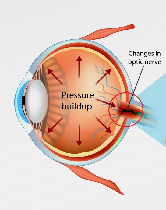
Glaucoma is a group of diseases that occur in the optic nerve of the eye. Most often, it is associated with elevated eye pressure. Over time it slowly damages nerve fibers in the optic nerve. Glaucoma is one of the leading causes of preventable blindness around the world!
Glaucoma in Austin

What Causes
Glaucoma?

The eye contains a drain called the trabecular meshwork. This drain system handles how fluid exits from inside the eye. In glaucoma, this drain becomes diseased. This causes fluid to build up and the pressure inside the eye to rise. Over time, the elevated pressure causes the optic nerve fibers to die. Once this occurs, it leads to irreversible vision loss.

What are the Symptoms of Glaucoma?

Unfortunately, most patients cannot tell if they have glaucoma. This is because glaucoma robs the eye of its extreme peripheral vision first. We are not as sensitive to this type of vision loss. Also, elevated pressure normally does not cause pain. This is why glaucoma testing on routine examination is so important. The only way the condition can be detected before any kind of visual impairment is through an eye exam. Acute angle-closure glaucoma is the only type that may exhibit early symptoms. These may include eye sensitivity to bright light, blurred vision, nausea, and eye pain.

Once glaucoma has developed, symptoms may include:

https://www.mediawebtool.com/mobile.php?cnr=SSO12-WESP&single=586&size=nor&log=schw

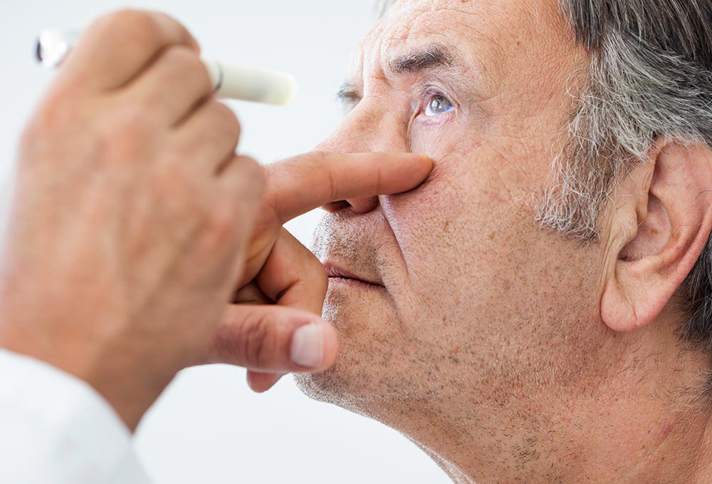
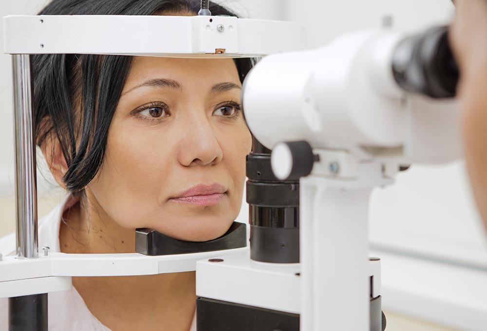
How is Glaucoma Diagnosed?

Tests used by ophthalmologists to diagnose glaucoma include:

  • Tonometry: Tonometry is a procedure that checks the intraocular pressure (IOP).
  • Pachymeter: A pachymeter test measures the thickness of the cornea. A thin cornea and high IOP show glaucoma or the possibility of developing it.
  • Gonioscopy: This is a test that looks at the anterior chamber, which is the front part of the eye. A gonioscopy checks whether the trabecular meshwork, where the aqueous humor drains out, is closed or open.
  • Optic Nerve Imaging: This test called the OCT helps to detect early nerve damage before vision loss.
  • Visual Field Testing: This test checks for any vision loss. It helps gauge the progress of glaucoma.

A comprehensive eye exam may involve most of these tests. This allows ophthalmologists at Westlake Eye Specialists to detect glaucoma at an early stage.

How is Glaucoma Treated?

There’s no cure for glaucoma. When diagnosed early on, it is treatable. The different types of treatments used for glaucoma are:

Durysta

Durysta is a new cutting edge injectable Glaucoma therapy now available in the US. It reduces Intraocular Pressure caused by Open-Angle Glaucoma by inserting a microscopic 1mm biodegradable implant into the eye. The implant contains the drug Bimatoprost, and once the implant is in the patient’s eye, it slowly releases this drug over time to reduce the patient’s eye pressure and decrease their reliance on topical drops. In two different studies, Durysta reduced intraocular pressure in patients with open-angle glaucoma by up to 30%. The implant lasts 4-6 months, but some ophthalmologists are reporting that their patients are continuing to experience lower pressure for 12-24 months after receiving the therapy. Durysta allows many patients to have another option for treatment who would otherwise suffer from dry eye related to drops, forget to take their medications or struggle to afford them. Westlake Eye is proud to be the leader in the use of this technology in Central Texas.

Eye Drops

Eye drops decrease the intraocular pressure in the eye. They relieve the pressure on the optic nerve, preventing further damage.
Types of glaucoma eye drops include beta-blockers prostaglandin analogs, alpha agonists, carbonic anhydrase inhibitors, and rho kinase inhibitors. These eye drops work in two ways:

  • They increase the rate of drainage of the aqueous humor from inside the eye
  • They decrease the rate of production of the aqueous humor

Eye Surgery

Glaucoma surgery is performed to lower pressure inside the eye. If eye drops are not effective, an ophthalmologist will recommend surgery. The types of glaucoma surgery include:
Looking into eyes

Minimally-Invasive Glaucoma Surgery (MIGS)

The procedure involves making a small incision in the eye. This leads to increased drainage of the aqueous humor and lowered intraocular pressure.
Omni

The OMNI Glaucoma Treatment System

Dr. Vendal is one of the first glaucoma specialists in Austin to use the OMNI system during MIGS for patients with open-angle glaucoma. The OMNI system helps reduce intraocular pressure and can be completed at the same time as cataract surgery. This is a revolutionary new treatment system, allowing patients with open-angle glaucoma to have the best possible results.
Hydrus

Hydrus Microstent

The Hydrus Microstent is another minimally invasive glaucoma procedure. With the Microstent, the less invasive approach means fewer complications and faster healing. The Hydrus Microstent is about the size of an eyelash and placed using microscopic incisions.
Gel Stent

The XEN® Gel Stent

The XEN® Gel Stent is a small tube that is surgically implanted into the eye and becomes soft and flexible. The implant is designed to lower high eye pressure in open-angle glaucoma patients. The XEN® Gel Stent is often recommended if previous surgical treatment has failed or other medications didn’t help. The XEN® Gel Stent creates a small channel in the eye to drain fluid and help lower eye pressure. The XEN® Gel Stent is very small—about the length of an eyelash. It is placed just under the conjunctiva, which is the clear membrane that covers the white of your eye. The XEN® Gel Stent stays in the eye permanently. You may or may not need to use glaucoma eye drops after the XEN® procedure.

Trabeculectomy

In this type of surgery, a small part of the sclera (outermost layer of the eye), is removed. As a result, an extra channel is created in the trabecular meshwork. This allows the aqueous humor to be drained into the area below the conjunctiva. Compared to MIGS procedures, a trabeculectomy is a more invasive procedure. This means that patients will experience a longer recovery period. Trabeculectomy can be combined and performed with cataract surgery at the same time. It can also be performed again on the same eye at a later time if necessary.

Ahmed Valve Implant

The Ahmed valve is a silicone drainage device that lowers eye pressure. It does this by allowing fluid to leave the inside of the eye through a tube. The valve is implanted onto the white part of the eye. Its silicone tip is then pierced through to the inside of the eye, and it is permanently sewn in. The tip is microscopic in size, clear in color, and sits in front of the iris. It is not visible to the naked eye. The inside of the tube has a valve in place so that fluid only exits the eye as it builds up. This surgery takes about an hour to undergo.

Before Surgery:

Patients undergo glaucoma surgery at an outpatient surgery center with mild sedation and a numbing injection to the eye which deadens all sensation. The eye is prepped with eye drops before surgery. If patients are on any blood-thinning medications, they are usually asked to stop them about 1 week before surgery.

After Surgery:

The eye is patched and shielded after the surgery is complete. This patch is then left in place until the next day and removed in the clinic. Patients then begin a series of drops to the eye that is slowly tapered off over 1 month. Vision returns over a period of 1-2 weeks and patients are seen up to 1-2 times a week if needed.

During visits, more anti-scarring medication is sometimes added to the eye. A contact lens may be placed on the eye as needed.

Patients are asked not to bend over, lift heavy things, or get the eye dirty during the healing period.

Laser Treatments

Light pulse lasers are used to lower eye pressure safely in the clinic. Types of laser treatments for glaucoma include:

Selective Laser Trabeculoplasty (SLT)

SLT is a frequently used laser treatment for open-angle glaucoma. The goal of the treatment is to help fluids drain out of the eye. It aims to reduce intraocular pressure that can cause damage to the optic nerve and loss of vision. The selective technique is much less traumatic to the eye than ALT. Before SLT, ALT was the standard laser procedure.

Laser Peripheral Iridotomy (LPI)

LPI is usually the procedure of choice when treating angle-closure glaucoma. It creates a hole in the iris’s outer edge, which then leads to an opening of the angle. By widening the angle, it helps improve fluid outflow. Rather than improving your vision, iridotomy is really meant to preserve remaining vision.

Cyclophotocoagulation

During laser cyclophotocoagulation, two different kinds of laser technology are used. In the first, a laser probe is placed on the white wall of the eye, and the top and bottom half of the eye is treated for 2 minutes. For the second, a telescopic device gets placed through a small incision at the cornea’s edge. This helps visualize the parts of the eye that make the ciliary body (internal fluid of the eye). Laser energy is then directed to this area. The two treatments work together to help your eye produce less fluid and lower intraocular pressure.

Are There Any Side Effects When Undergoing Glaucoma Treatments?

Eye drops may cause itching, redness, burning sensation, drowsiness, and headaches. Glaucoma surgery may have side-effects such as cataracts, low intraocular pressure, and even result in vision loss. More temporary side effects of surgery include eye swelling and soreness.

Is Glaucoma Preventable?

There are ways to lower the risk of glaucoma or possibly prevent it. High pressure is a risk factor for glaucoma. Other risk factors for glaucoma are:

Normal-Tension Glaucoma Risk Factors

  • Japanese ethnicity
  • Family history of glaucoma
  • Cardiovascular disease
  • Autoimmune Disease
Eye

Open-Angle Glaucoma Risk Factors

  • Family history of glaucoma
  • Thin corneas
  • High pressure in the eyes
  • The suspicious appearance of the optic nerve with increased cupping
  • 60 years and older for the general population or Mexican Americans
  • Being 40 years and older for African Americans
  • Use of corticosteroids such as creams, eye drops, inhalers, and pills
  • Eye injury or surgery
  • Diabetes
Getting Eyes Checked

Angle-Closure Glaucoma Risk Factors

  • Inuit and East Asian ethnicity
  • Family history of glaucoma
  • Eye surgery or eye injury
  • Farsightedness
  • 40 years and above

Lowering the Risk of Glaucoma

If you are at risk of developing glaucoma, take up a healthy lifestyle. This includes a nutritious diet and regular physical exercise. It’s also important to take care of your emotional and mental health. For your physical well-being, you should:

  • Avoid smoking
  • Maintain a healthy weight
  • Exercise daily
  • Reduce caffeine intake
  • Control medical conditions such as high blood pressure
  • Go for frequent, comprehensive eye exams


Glaucoma is one of the leading causes of blindness in the world. One of your best lines of defense is having regular eye exams. Our eye doctors specialize in the diagnosis and surgical management of glaucoma.

Who Is At Risk of Getting Glaucoma?

Anyone can get glaucoma. The risk of developing glaucoma only increases with age. Other risk factors include genetics and physical trauma to the eye. People with diabetes are also at a higher risk of developing glaucoma.

Glaucoma Treatment in Austin, Texas

If you can’t remember the last time you had an eye exam, it’s already been too long! Don’t let glaucoma steal your sight. Schedule an appointment with the experts at Westlake Eye Specialists in Austin, TX!

Request A
Consultation

To request a consultation with Westlake Eye Specialists, please click link below.